MRI Brain Scan Centre in Delhi
Brain MRI Scan: Test, Benefits, Procedure & Cost
Starting only @
Brain MRI SCAN
Book Your Brain MRI Scan Online at the Best Price in Delhi/NCR with Carebox.
Schedule your scan with ease through Carebox at trusted NABL & NABH accredited diagnostic centers. Call Now At +91 99536 30773 to Book your MRI Scan at 50% Discount.
Exclusive Benefits with Carebox

Personalized support to help you find the best centers nearby

Guaranteed discount on your scan at any center you choose
Get up to 60% additional discount on All tests & Scans
Skip the wait—book priority appointments for your scans.

Why CareBox is the Right Choice for You
Over 1.5 Lakh Patients Trust Us for Their Tests Every Month

Get Up to 60% Off on Every Scan—Start Saving Today!

100% Reliable and Accurate Reports

ISO and NABH Certified Scan Center
Brain MRI, also known as Magnetic Resonance Imaging of the Brain, Cranial MRI, and Neuroimaging. A brain MRI (Magnetic Resonance Imaging) scan is a medical imaging test that employs powerful magnets and radio waves to generate detailed pictures of the brain and the surrounding tissues. It allows physicians to visualize the structure of the brain and detect most abnormalities or conditions.
What Are the Benefits of Brain MRI?
There can be certain definite benefits that can be assigned to brain MRI in comparison to other imaging technology:
Brain MRI is very sensitive rate-wise since it contains two magnetic fields:
- High-Resolution Images: MRI takes high-resolution images of the brain’s soft tissues, i.e., cerebrospinal fluid, blood vessels, gray matter, and white matter. MRI gives good visualization even for small abnormalities.
- No Ionizing Radiation: While CT scans and X-rays use no ionizing radiation, MRIs neither use any ionizing radiation and are thus safer if a repeat serial scan is on a repeat process or for sensitive cases like women in pregnancy (although first trimester pregnancy MRIs are now also being avoided unless strictly unavoidable in futility cases).
- Multiplanar Imaging: MRI is capable of imaging more than one plane (axial, sagittal, coronal, and oblique) without having to restage the patient. Gives a general impression of the brain in very dissimilar orientations.
- Excellent Soft Tissue Contrast: MRI can distinguish contrast better between different soft tissues and is thus extremely sensitive to processes like plaques of multiple sclerosis, brain tumor, stroke, and infection.
- Functional MRI (fMRI): The most up-to-date of the MRI techniques, fMRI is able to quantitate alterations in the pattern of blood flow and thus can measure brain function. It is especially well-suited for studies and for generating maps of brain functional regions prior to surgery.
- MR Angiography (MRA) and MR Venography (MRV): Both of these MRI scans can visualize brain blood vessels without intra-arterial injection or other internal imaging like conventional angiography. MRA visualizes arteries, and MRV visualizes veins.
What Are the Pre-Procedure Steps Before a Brain MRI?
A brain MRI scan prep is low but regularly required:
- Notify Your Doctor: Tell your doctor about any illness, such as kidney disease (as this is most likely to impact the use of contrast media), or allergy, pregnancy, or lactation, and other medical devices implanted, such as pacemakers, defibrillators, cochlear implants, or neurostimulators. There are a few with no other choice for MRI compatibility or other special considerations.
- Medication: You can continue with your regular daily medications unless otherwise instructed by your doctor.
- Fasting: You will not usually need to fast before a brain MRI unless you are having contrast. If you are having contrast, your doctor has asked you to have food and liquid restricted for you.
- Clothing: Loose-fitting, comfortable clothing without metal clasp closures. You will be provided with a hospital gown.
- Metallic accessories should be prohibited: Do not wear any of the metal items such as jewelry, watches, credit cards, keys, hairpins, or any other metal items during scanning or remove them prior to scanning. Metallic equipment warps the magnetic field and leads to image distortion.
- Claustrophobia: If you suffer from claustrophobia (don’t want to be enclosed in a small space), notify your physician in advance. Your physician can provide you with a light sedative so that you’ll be more comfortable to have testing done. They have open MRI units that are less restricting, but the image isn’t going to be such a great photo with standard MRI equipment.
- Bring Past Scans: If you had previous earlier scans of your brain (MRI or CT), please do bring those with you on the appointment day. The radiologist could then review the news with previous scans.
- Follow Special Instructions: Always comply with special instructions of your doctor or imaging facility.
What to Expect During a Brain MRI Scan?
The brain MRI scan process utilizes a very powerful magnetic field, radio waves, and a computer to create highly precise images of brain tissues. MRI has no relation to any ionizing radiations like X-rays and CT scans. It is performed step by step, following below in detail:
- Preparation: You will usually have a questionnaire to complete regarding your disease history, implants (that is, metal devices such as pacemakers), and allergies prior to testing. You’ll be asked to wear a hospital gown and remove all your jewelry, watch, hearing aid, and other metallic objects that will interfere with the magnetic field.
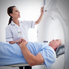
- Position: You will lie on a moving table that is inside the MRI machine. The machine is huge, tube-like, and open on both ends. Pillows and straps can be around you to hold you in position. Your head will be inside a special coil, a type of equipment that sends and receives radio waves, when having a brain MRI, and the brain image will thereby be enhanced.
- Image Acquisition: Next, the technologist will proceed to a control room, usually in close vicinity. You can talk to them via an intercom. The scanner will initiate with extremely loud thudding, tapping, or humming noises. Don’t get alarmed by that; don’t get frightened due to reversing magnetic field gradients. Try to remain as motionless as possible for that duration, as movement will lead to image blurring. Scanning is typically a series of pictures over several minutes or so per station. There are many series that picture many different brain tissue structures or picture many different abnormalities.
- Contrast Agent (Optional): Intravascular contrast agent administration (usually gadolinium-based) is allowed on a selective intra-procedure basis. Contrast agents will be used to outline discrete structures, vessels, or fields of tumor or inflammation. The decision whether to use contrast is left to the radiologist and clinical question.
- Scan Completion: Once your images have been scanned, the table will come out of the scanner. Your images will be scan-checked by the technologist to make sure they are good quality before releasing you. Your result will be interpreted by a specially trained medical imaging doctor, and they will report your result.
How to Get a Brain MRI Scan at the Lowest Cost in Delhi?
You can have your brain MRI scan at the lowest cost in Delhi with the help of CareBox. We will help you to find the best imaging center for brain MRI near you with highly equipped machines, transparency of costs with no hidden charges, experienced radiologists, and all.
How to Book a Brain MRI at a CareBox?
To make an appointment for a brain MRI with the care box, you need to take care of all these things:
- Contact CareBox: Obtain Delhi’s best diagnostic imaging centers through CareBox.
- Ask if appointments can be booked: Call or email CareBox and inquire if brain MRI appointments can be scheduled at any imaging center near you. Specify a doctor’s referral and if you have any special requirements (e.g., require contrast, claustrophobia problem).
- Discuss the Cost and Preparation: Please ask how much you are going to be charged for the brain MRI scan and whether the radiologist fee and/or any other fee is included or not. Second, please ask for any special preparation instructions that you should follow prior to your visit to your appointment.
- Select Date and Time: Select a proper date and time for your brain MRI from your times and center times.
- Provide Your Own Information: You may be requested to give your own information, telephone numbers, and your history.
- Confirmation: Do ensure you have a date, time, center location, and instruction confirmation.
- Pre-Scan Preparation: Do the preps as instructed by the care box before their arrival visit to your appointment.
- Attend Your Visit: Bring along your documents at the center on time. Documents that are needed:
- Doctor’s prescription slip
- Documents related to medical history
- Any valid ID proof for verification
What Does the MRI of the Brain Show?
Brain MRI is a suitable imaging diagnostic agent for the observation of brain structure and identification of various types of abnormalities. Brain MRI can be used to diagnose and screen for any neurologic disorder.
Brain MRI can diagnose especially:
- Brain Tumors: MRI is sensitive enough to differentiate malignant and benign tumors and size, extent, and effect on nearby brain tissue.
- Stroke: MRI will be able to differentiate between brain damage areas due to stroke-like ischemic (obstructed flow) and hemorrhagic (bleeding) stroke. Diffusion-weighted imaging (DWI), a form of MRI sequence, is found useful in acute stroke initial evaluation.
- Multiple Sclerosis (MS): MRI will be able to show typical brain and spinal cord plaques or MS lesions. MRI is utilized for diagnosis, disease activity, and evaluation of response to therapy.
- Infection: Brain inflammation (encephalitis), meninges covering brain and spinal cord inflammation (meningitis), and brain abscess can be diagnosed on brain MRI.
- Hemorrhage (Bleeding): Traumatic intracranial hemorrhage, rupture of an aneurysm, or otherwise can be outlined in an MRI. Depending on the MRI sequences employed, the amount of bleeding can be identified.
- Hydrocephalus: Ventricular or cerebrospinal fluid space dilatation secondary to cerebrospinal fluid accumulation can be seen on MRI.
- Developmental Abnormalities: MRI detects congenital maldevelopment of the brain.
- Traumatic Brain Injury (TBI): MRI will technically confirm brain trauma, i.e., hematomas, contusions, and diffuse axonal injury, but CT scanning is employed as a first-line imaging in head trauma.
- Dementia and Alzheimer’s Disease: MRI will exclude other causes of intellectual loss and will demonstrate normal patterns of brain atrophy in the dementias and Alzheimer’s disease.
- Seizure Disorders (Epilepsy): MRI will detect structural brain lesions that cause seizures, e.g., hippocampal sclerosis or tumor.
- Vascular Abnormalities: MRA will detect arteries and aneurysms, arteriovenous malformations (AVMs), and stenosis (an artery narrowing). MRV can see the veins to diagnose thrombosis (blood clots).
- Disorders of the Pituitary Gland: MRI perhaps can see the pituitary gland and nearby structures very well for tumor or other endocrine disease hormone secretion diagnosis.
- Complications and Sinusitis: MRI is not the first-line drug in sinus staging, but in some situations it can be used in complicated sinus infection evaluation or intracranial spread of such infection.
How Long Will the MRI of the Brain Take?
- A brain MRI scan length will depend on many things changing, such as
- Clinical indication: If the scan is indicated, then this will be a sign of how many image sequences will be needed and what kind.
- Use or lack of contrast: A contrast brain MRI will take longer compared to when contrast is not used since one is time-consuming when putting in and passing contrast.
- Number of sequences ordered: More complex cases or more scanning of specific areas will take longer sequences and therefore longer to scan.
- MRI-type machine: More advanced and quicker machines will be faster.
- Specialized techniques employed, such as fMRI, MRA, or MRV, will contribute to total scan time.
What are the Risks and Side Effects of a Brain MRI?
A brain MRI is an extremely safe test for the fact that it has nothing to do with ionizing radiation. There are some risks and side effects, yes, as one quotes:
Magnetic Field Hazards: The intense magnetic field used in MRI is hazardous to patients with some metal implants or foreign bodies in their body. Inform your doctor and MRI technologist in advance about such implants, if any, for example:
Pacemakers and implantable cardioverter-defibrillators (ICDs) (they can be MRI-conditional or MRI-safe, but that needs to be checked).
- Cochlear implants.
- Some aneurysm clips.
- Metallic heart valves.
- Implanted drug infusion pumps.
- Neurostimulators.
Metallic foreign bodies (e.g., from prior trauma). These can short out, become lost, or heat up when inserted into the magnetic field and thus cause disastrous damage.
- Contrast Agent Reactions: Alkali is uncommon with gadolinium contrast agents. Any reaction of any sort that does occur usually is in its mildest form, i.e., hives or pruritus, but it occurs sparingly. Alkali, especially contrast agent alkali, must be brought to the physician’s notice.
- Nephrogenic Systemic Fibrosis (NSF): Gadolinium-based contrast agents have been associated with a benign but potentially toxic process, nephrogenic systemic fibrosis (NSF), in the acutely ill kidney patient. It should be disclosed to the physician if there is any kidney abnormality prior to administering contrast. Physicians typically screen renal function prior to using gadolinium-based contrast.
- Claustrophobia: Claustrophobia, or the fear of confinement within small spaces, may cause some patients to feel anxious or uncomfortable in the same room where an MRI scanner is installed. It is a state of mind. Open scanners, as mentioned earlier, or sedation can be used for this purpose.
- Noise Exposure: The worst that will happen is the momentarily deafening, ear-shattering, piercing ringing of the MRI equipment for a moment or, in the extremely remote chance, temporary hearing loss with no ear protection. Headsets or earplugs are always used to make this a nonissue.
- Burns: Burns are unlikely but may occur if metal items come in contact with the skin during the MRI procedure. Hence, ornaments must be removed, and dressing must be removed carefully.
When Will I Receive My Brain MRI Report?
How and when you receive your brain MRI report will vary by imaging center and by report level. But here is the typical pattern, and you will go through something like that:
- Technologist Initial Review: After the scan has been completed, the MRI technologist will typically review the images to determine if they are appropriate for quality and if all of the requested sequences have been acquired.
- Radiologist Interpretation: Then, they are interpreted by a board-certified physician radiologist who will take careful readings of the images and interpret them. Yes, it does take a while because the radiologist will have to interpret the images and cross-match them with the clinical information your referring physician has provided.
- Report Generation: There will be a written report of findings generated by the radiologist. The report is a catalog of structures seen in the brain and any abnormality detected.
- Report to Your Doctor: Your final radiology report typically will be reported to your referring physician in a timely fashion. This is 24 to 48 hours for simple cases. For some urgent cases, the radiologist will report earlier to your physician with important findings.
- Category: Radiology Reports.
- Follow-up Visit with Your Doctor: You might get a follow-up visit to visit your referring doctor to get your brain MRI reports. Your doctor will place findings in the perspective of your symptoms and history and make a value call on what you should do next, i.e., treatment, further studies, or watch out..RI
How to Book Brain MRI Scan At Affordable Cost
Call us or chat with our customer service agent.
We will answer all your question and help to book your appointment.
Lab details & charges will be shared with you over the call.
We’ll book your test as per your preferred date and time slot.
Call us or chat with our customer service agent.
We will answer all your question and help to book your appointment.
Lab details & charges will be shared with you over the call.
We’ll book your test as per your preferred date and time slot.