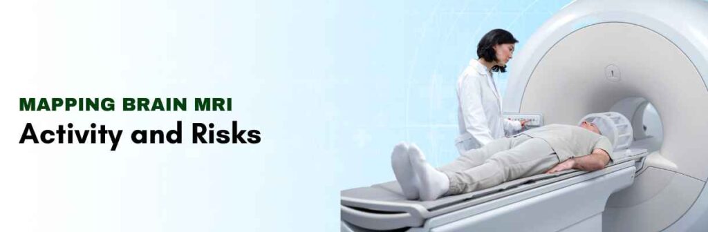
A brain MRI (magnetic resonance imaging) scan, or head MRI, is a painless test that creates very detailed pictures of the structures within your head — primarily, your brain. It doesn’t involve radiation.
Mapping Brain Activity
When a part of the brain is activated, its neurons need higher levels of oxygen. The blood flow to supply the needs of that tissue will increase – thus providing the where and when of oxygenated blood flow.
Blood oxygenation level dependent (BOLD) contrast is the main mode of FMRI. BOLD is a form of FMRI signal that exists because of the differences in magnetic properties of oxygenated and deoxygenated hemoglobin.
Oxygenated hemoglobin is diamagnetic and deoxygenated hemoglobin is paramagnetic (meaning that in a magnetic field they are interacted with locally differently producing measurable differences in signal intensity from an MRI scanner). More neural activity will produce greater BOLD signals overall because more neural activity will result in higher levels of blood flow and higher levels of blood oxygen.
While participants are being scanned with an FMRI, they may be asked to perform (e.g., moving a finger, reading, or listening to sounds). These tasks are typically simple, actions or thoughts that will cause varying activity in the brain. The scanner will record a series of fast, repeated still images of the brain.
Specialized software will then examine each of the images to determine the changes to blood flow and oxygenation to different areas of the brain associated with the task. The images with all of that data are then processed into activation maps – color-coded images that include the most active brain areas associated with certain behaviors, tasks, cognitive processes, or tasks.
Risks of Brain MRI
While we typically presume that brain MRIs are safe imaging techniques without ionizing radiation exposure, there are safety issues:
The magnetic field can move or heat metal im-plants or surgical pins.
Due to the risk of allergic reaction with some patients, contrast material can be a problem in the case of contrast MRI. Patients can become uncomfortable due to the intense noise produced by the MRI scanner. Patients can be anxious due to being in a small space, especially if they have claustrophobia about the enclosure of the MRI scanner.
It is advised for women to avoid breastfeeding for a timely period (the approx. 48 hours) after contrast is given to the breastfeeding mother.
Conclusion
Overall, a brain MRI is an excellent diagnostic imaging modality that is safe, noninvasive and provides high-resolution images of brain structure and function. Functional MRI (fMRI) allows for further maps of brain activity by monitoring blood flow and the oxygenation of hemoglobin based on neuronal activity.
There are inherent risks from an MRI, including the inherent discomfort from the noise of unbearable decibels, claustrophobia, metal implants, or side effects from contrast agents that will be considered. As a whole, the brain MRI is an invaluable diagnostic technique that can inform patient assessments of brain abnormalities, support therapy options, and help us understand how the brain functions when healthy and diseased.
Frequently Asked Questions
Q. What is a contrast MRI brain scan?
A contrast MRI brain scan involves a contrast agent which enhances the quality of the brain images provided in the MRI scan allowing changes and abnormalities to be seen more easily.
Q. Can MRI scans detect brain cancer?
Yes, MRI scans are especially adept at detecting tumors including brain cancer.
Q. Where can I have a brain MRI in Delhi?
You can get the best diagnostic centre in Delhi via Carebox, where you can book best brain MRI scan.
Q. What are MRI and FMRI used for?
MRI is used to provide high-resolution pictures of the internal structures of the body while FMRI is a specialized MRI that measures brain activity and maps it by changes in blood flow and then oxygenation.
Q.What are the clinical applications of functional brain MRI?
Functional brain MRI (FMRI) has many beneficial clinical applications in many areas, particularly for neurosurgical preoperative planning for surgical resection (tumors, vascular malformations, etc.), planning surgery for patients with epilepsy (focal seizures), and helping to define brain functions, in patients with possible different types of neurological disease.
